Research area

Brain Metastases

Where do brain metastases arise, does it depend on the primary cancer, and can whole brain radiotherapy spare the regions where they rarely occur without missing disease?

NIHNIH/NCI R01CA292043 · 2025–2030 · Development of a Personalized Voxel-wise Prediction of Brain Metastases using Multi-Parametric MR Imaging to Reduce Treatment ToxicityNIH RePORTER ↗

Approach

From many patients to one map

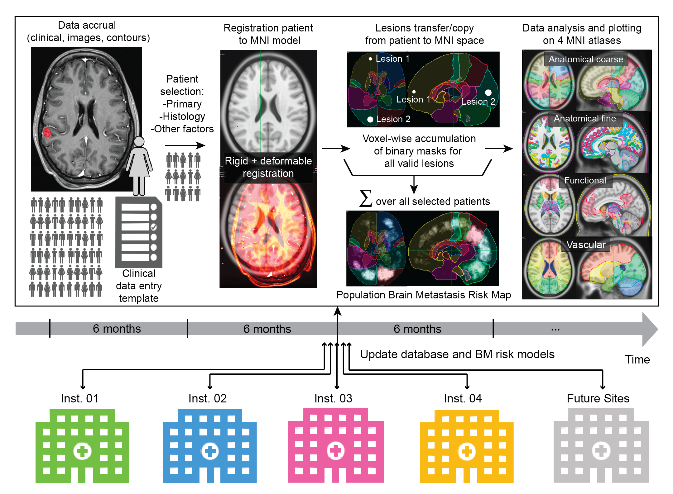
  1. 01De-identify. HIPAA Safe Harbor, with structure names checked against AAPM TG-263.
  2. 02Register. Rigid then deformable registration of each patient to MNI152.
  3. 03Accumulate. Lesion masks summed voxel-wise across patients, by primary cancer.
  4. 04Analyze. Frequencies reported on coarse and fine anatomical, functional and vascular atlases.
Atlas workflow from data accrual to analysis on four reference atlases
Fig. 1 Atlas workflow, updated as institutions contribute.1

Results

What the atlas shows

  • Metastases localize predominantly to high-perfusion regions near the grey and white matter junction.1
  • Lung and breast cancers, in contrast to melanoma, frequently metastasize to the cerebellum.1
  • Deep brain structures are relatively spared regardless of primary cancer, the basis for sparing them in whole brain radiotherapy.1
Interactive resourceBrain AtlasExplore the 13,926 lesions of the current update by primary cancer and reference atlas, in three planes and in 3D.Open the Brain Atlas
MethodDeep Learning Auto-SegmentationSegmenting brain structures and lesions across MR and CT with little expert annotation.Open

Status

WBRT PROTECT: aims and milestones

Whole-Brain RT: Personalized Radiation Optimization To Eliminate Collateral Toxicity. Detailed reports are shared with collaborators after sign in.

Year 1Year 2Year 3Year 4Year 5
Aim 1 · Personalized risk model
Population risk mapComplete
Anatomy, perfusion, historyIn progress
Predictive evaluationPlanned
Aim 2 · Dosimetric impact
In-silico proof of conceptComplete
30-patient planning studyIn progress
Aim 3 · Clinical evaluation
Protocol and regulatoryIn progress
In-silico trial, 3 sitesPlanned
Phase I, 40 patientsPlanned

Collaboration

Contributing institutions

4 institutions in the United States and Canada. Co-authors of the Nature Communications atlas (reference 1), by affiliation. Patient numbers are those of the current atlas update.

University of California, San FranciscoRadiation Oncology · Radiology and Biomedical Imaging · Neurological Surgery · Epidemiology and Biostatistics · Pathology
1,762patients

Lead site. Imaging and contours, registration to MNI space, accumulation and analysis.

Jorge Barrios, Evan Porter, Dante Capaldi, Taman Upadhaya, William C. Chen, Javier E. Villanueva-Meyer, Gilmer Valdes, Fei Jiang, Hui Lin, Oi Wai Chau, Benjamin Ziemer, Maasa Seaberg, Penny K. Sneed, Jean L. Nakamura, Lauren C. Boreta, Shannon E. Fogh, David R. Raleigh, Jessica Chew, Harish Vasudevan, Soonmee Cha, Christopher Hess, Shawn L. Hervey-Jumper, Philip Theodosopoulos, Daniel Cooke, Ke Sheng, Steve Braunstein, Olivier Morin

Princess Margaret Cancer CentreRadiation Medicine Program, University Health Network · Medical Biophysics, University of Toronto
539patients

MRI and contours, processed with the same pipeline as UCSF.

Michael Maddalena, David B. Shultz, Derek S. Tsang, Jan Seuntjens, Catherine Coolens

University of California, DavisRadiation Oncology, UC Davis Health
479patients

MRI and contours, processed with the same pipeline as UCSF.

Julian R. Perks, Payton H. Stone, Jinger Y. Sun, Muhtada A. K. Aldin, Ruben Fragoso, Stanley H. Benedict

Memorial Sloan Kettering Cancer CenterRadiation Oncology · Medical Physics
510patients

Contours already mapped to MNI space at Memorial Sloan Kettering.

Aditya Apte, Michalis Aristophanous, Eve LoCastro, Dylan Hsu, Ase Ballangrud, Kayla Prezelski, Luke Pike, Joseph O. Deasy


Publications

Papers in this area (3)

  1. 2025
    Barrios J, Porter E, Capaldi DPI, Upadhaya T, Chen WC, Perks JR, Apte A, Aristophanous M, LoCastro E, Hsu D, Stone PH, Villanueva-Meyer JE, Valdes G, Jiang F, Maddalena M, Ballangrud A, Prezelski K, Lin H, Sun JY, Aldin MAK, Chau OW, Ziemer B, Seaberg M, Sneed PK, Nakamura JL, Boreta LC, Fogh SE, Raleigh DR, Chew J, Vasudevan H, Cha S, Hess C, Fragoso R, Shultz DB, Pike L, Hervey-Jumper SL, Tsang DS, Theodosopoulos P, Cooke D, Benedict SH, Sheng K, Seuntjens J, Coolens C, Deasy JO, Braunstein S, Morin O. Multi-institutional atlas of brain metastases informs spatial modeling for precision imaging and personalized therapy. Nat Commun. 2025 May 15; 16(1):4536. PMID: 40374598. PMCID: PMC12081687PMID 40374598
  2. 2024
    Salans M, Ni L, Morin O, Ziemer B, Capaldi DPI, Raleigh DR, Vasudevan HN, Chew J, Nakamura J, Sneed PK, Boreta L, Villanueva-Meyer JE, Theodosopoulos P, Braunstein S. Adverse radiation effect versus tumor progression following stereotactic radiosurgery for brain metastases: Implications of radiologic uncertainty. J Neurooncol. 2024 Feb; 166(3):535-546. PMID: 38316705. PMCID: PMC10876820PMID 38316705
  3. 2022
    Sneed PK, Chan JW, Ma L, Braunstein SE, Theodosopoulos PV, Fogh SE, Nakamura JL, Boreta L, Raleigh DR, Ziemer BP, Morin O, Hervey-Jumper SL, McDermott MW. Adverse radiation effect and freedom from progression following repeat stereotactic radiosurgery for brain metastases. J Neurosurg. 2022 May 20; 1-9. PMID: 35594891PMID 35594891

Updates

News from this area

  1. 2025-08Brain MetastasesWBRT PROTECT (R01CA292043) funded by the National Cancer Institute.
  2. 2025-05Brain MetastasesMulti-institutional brain metastasis atlas published in Nature Communications.

References

  1. Barrios J, Porter E, Capaldi DPI, et al. Multi-institutional atlas of brain metastases informs spatial modeling for precision imaging and personalized therapy. Nat Commun. 2025;16:4536. doi:10.1038/s41467-025-59584-7
  2. Morin O, Vallières M, Braunstein S, et al. An artificial intelligence framework integrating longitudinal electronic health records with real-world data enables continuous pan-cancer prognostication. Nat Cancer. 2021;2(7):709-722. PMID 35121948