
Method
Deep Learning Auto-Segmentation
Atlases, dose planning and quality control all start from contours. Can deep learning segment brain structures and lesions reliably across MR and CT, with little expert annotation?
Study
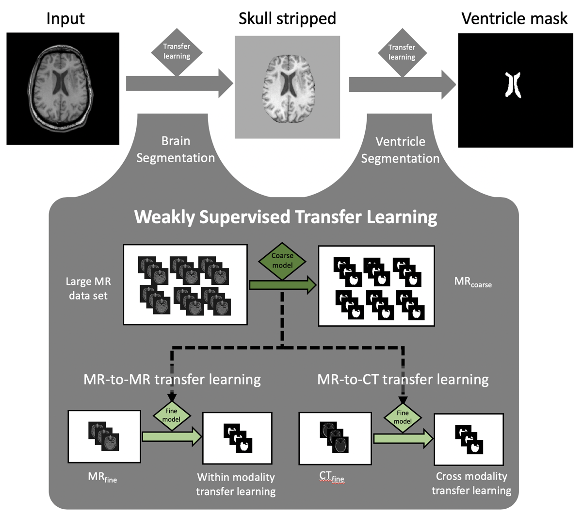
Weakly supervised transfer learning for multi-modality brain and ventricle segmentation
Retrospective, IRB-approved study of head MR and CT images. Datasets of 2,193, 162 and 160 images with coarse or fine annotations were used for training, validation and testing. Performance was measured with the Dice score and percent volume difference.
- Ventricle models trained with fine-grained data improved Dice from 0.75 to 0.82 and percent volume difference from 21.5% to 8.0% over coarse-grained models.
- Transfer learning improved Dice over training without it (0.86 vs 0.83, p < 0.01).
- Transfer learning models trained with as few as 20 images matched models trained with 100.
Turgutlu KC, Barrios Ginart J, … Villanueva-Meyer J, Morin O. Manuscript prepared for Radiology: Artificial Intelligence [status to confirm].
Data available: MR and CT, simple and complex annotation sets.
In use
Where segmentation runs today
Brain metastasis atlasBrain extraction with a quality gate before every patient is registered to MNI space.Brain Metastases
SCALE ConsoleThe same brain extraction runs at participating sites, inside a container.SCALE Console
Brain metastasis detection and segmentation[Project to add]